
When Do Doctors Recommend a CT Scan for Lung Disorders?
Breathing is something most of us rarely think about—until it becomes difficult. Whether it is a persistent cough, unexplained chest pain, shortness of breath, or recurrent lung infections, respiratory symptoms can significantly affect daily life and may indicate an underlying medical condition that requires prompt evaluation.
Lung diseases remain one of the leading causes of illness worldwide. Conditions such as pneumonia, tuberculosis (TB), chronic obstructive pulmonary disease (COPD), asthma, interstitial lung disease, pulmonary fibrosis, and lung cancer affect millions of people every year. Early diagnosis plays a crucial role in improving treatment outcomes, reducing complications, and enhancing quality of life.
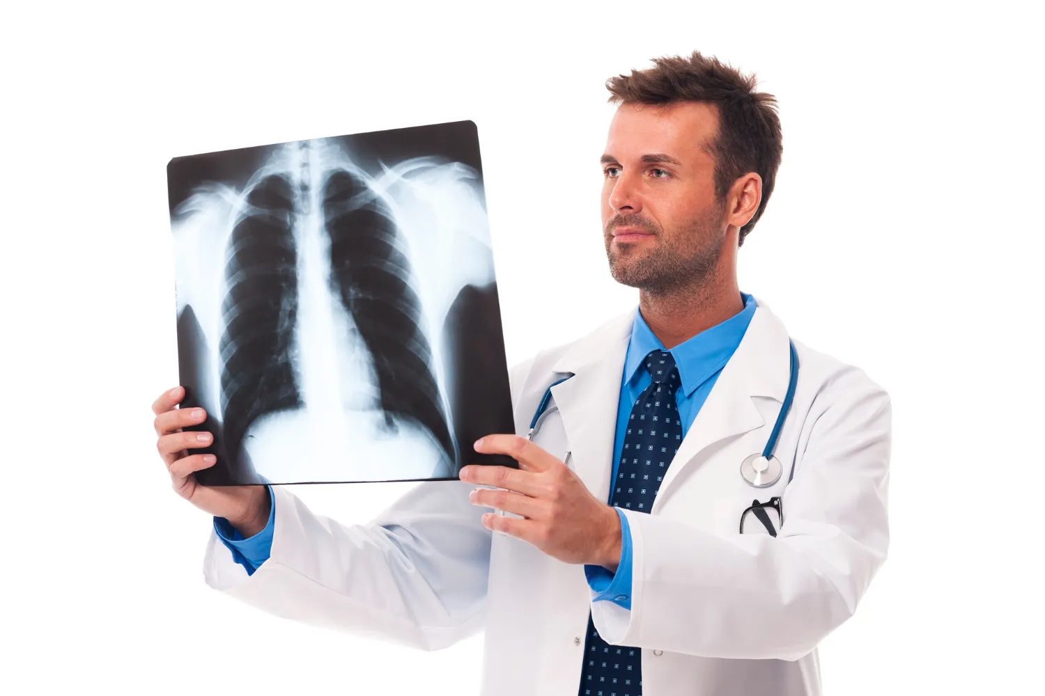
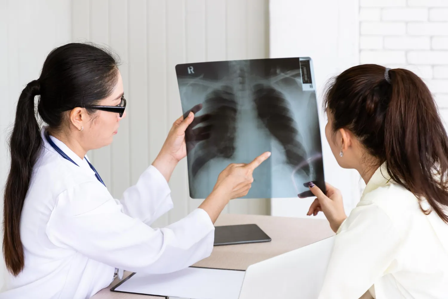
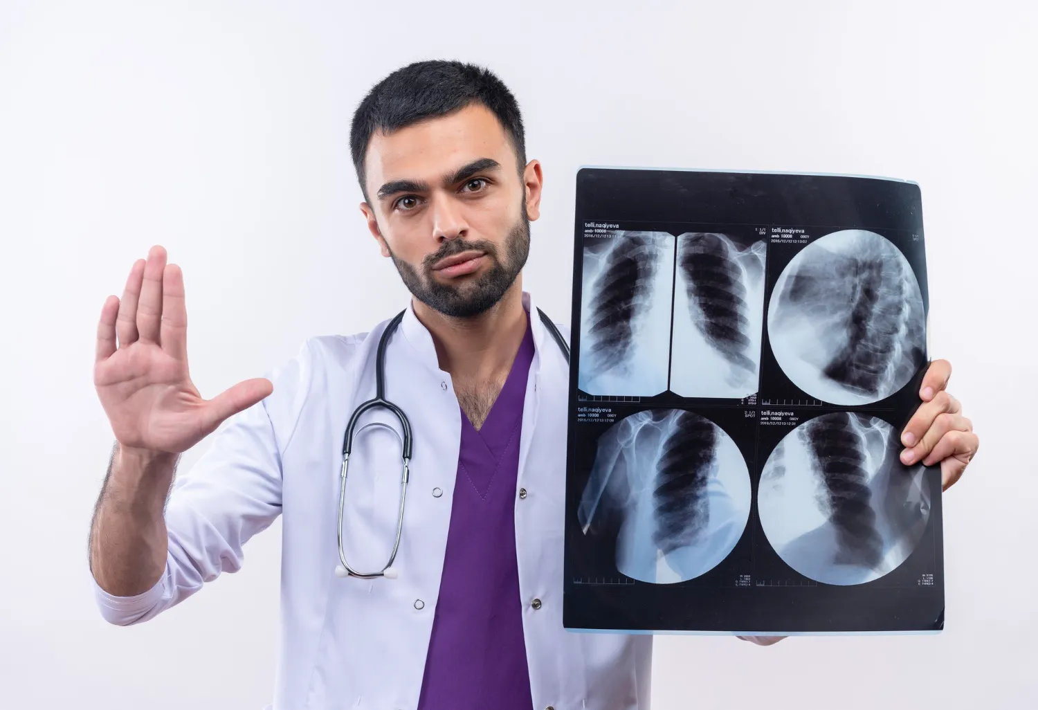
While a chest X-ray is often the first imaging test performed for respiratory symptoms, it may not always provide enough detail to identify the exact cause of the problem. In such situations, doctors frequently recommend a Computed Tomography (CT) scan of the chest, an advanced imaging investigation that produces detailed cross-sectional images of the lungs, airways, blood vessels, and surrounding structures.
Many patients wonder:
- Why do I need a CT scan instead of an X-ray?
- Does a CT scan detect lung infections?
- Can it identify lung cancer at an early stage?
- Is the procedure safe?
- Will I need contrast injection?
This comprehensive guide explains when doctors recommend a CT scan for lung disorders, how the procedure works, what conditions it helps diagnose, and how the results support better treatment decisions.
What Is a CT Scan?
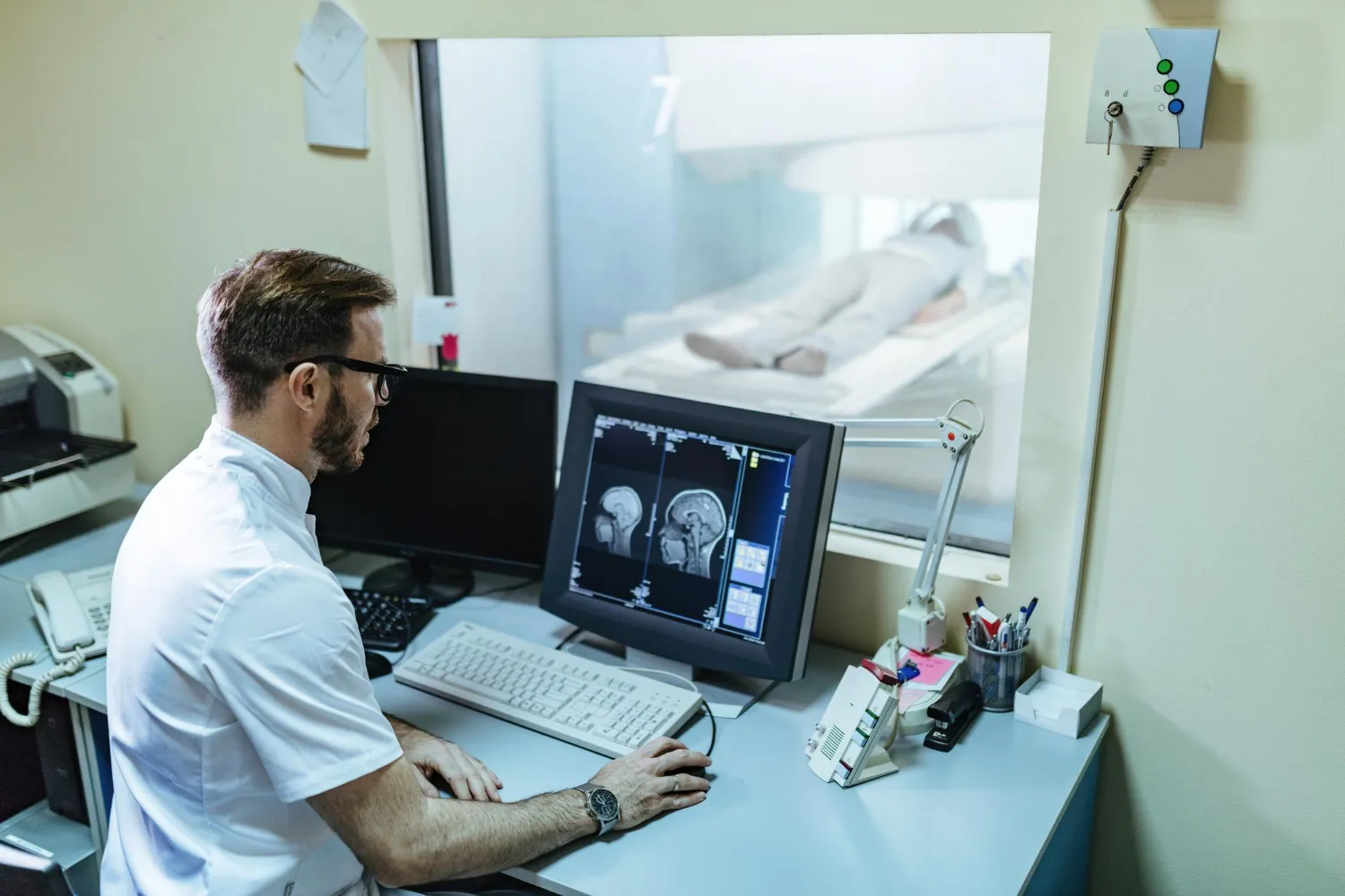
A Computed Tomography (CT) scan is an advanced diagnostic imaging technique that combines X-ray technology with sophisticated computer processing to create detailed cross-sectional images of the body.
Unlike a conventional chest X-ray, which produces a two-dimensional image, a CT scan captures multiple thin slices from different angles. These images are then reconstructed into highly detailed views of the lungs and surrounding tissues.
Because of its superior image quality, a CT scan can identify abnormalities that may not be visible on standard radiographs.
Why Is a CT Scan Better Than a Chest X-ray for Certain Lung Conditions?
A chest X-ray remains an excellent first-line investigation for many respiratory conditions. However, some lung diseases develop in areas that are difficult to visualize or are too small to detect using routine X-ray imaging.
A CT scan provides significantly greater detail and allows doctors to evaluate:
- Lung tissue
- Airways (bronchi)
- Blood vessels
- Pleura (lining around the lungs)
- Lymph nodes
- Mediastinum (central chest structures)
- Chest wall
This additional information often leads to earlier diagnosis and more accurate treatment planning.
When Do Doctors Recommend a CT Scan for Lung Disorders?
A CT scan is recommended when symptoms, physical examination findings, or previous investigations suggest that more detailed imaging is required.
Below are some of the most common situations where your doctor may advise a CT scan.
Persistent or Unexplained Cough
A cough lasting longer than eight weeks should never be ignored.
Although many persistent coughs result from allergies, asthma, acid reflux, or chronic infections, prolonged symptoms may occasionally indicate more serious underlying conditions.
A CT scan helps evaluate:
- Hidden infections
- Lung nodules
- Bronchiectasis
- Early lung cancer
- Chronic inflammatory lung disease
Detailed imaging enables doctors to identify the underlying cause more accurately than a routine chest X-ray alone.
Suspected Lung Infection
While pneumonia is commonly diagnosed using a chest X-ray, certain infections require more advanced imaging.
Doctors may recommend a CT scan if:
- Symptoms fail to improve with treatment.
- The infection keeps returning.
- The diagnosis remains uncertain.
- Lung abscess is suspected.
- Complications such as pleural collections are possible.
CT imaging provides detailed information about the location, severity, and extent of infection.
Tuberculosis (TB)
India continues to have one of the highest burdens of tuberculosis worldwide.
Although sputum testing remains essential for diagnosis, CT imaging often helps assess:
- Disease extent
- Cavitary lesions
- Lymph node involvement
- Complications
- Response to treatment
In patients with persistent symptoms despite treatment, CT scanning offers valuable additional information.
Evaluation of Lung Nodules
A lung nodule is a small round shadow seen within lung tissue.
Most nodules are non-cancerous and may result from previous infections or inflammation.
However, some require further evaluation because they may represent early lung cancer.
CT scanning helps doctors assess:
- Nodule size
- Shape
- Margins
- Growth over time
- Internal characteristics
These features help determine whether observation, biopsy, or further imaging is appropriate.
Suspected Lung Cancer
Early diagnosis significantly improves treatment outcomes in lung cancer.
Doctors frequently recommend CT scanning for patients who have:
- Persistent cough
- Blood in sputum
- Unexplained weight loss
- Persistent chest pain
- Long-term smoking history
- Abnormal chest X-ray findings
CT imaging assists in detecting lung tumours, evaluating their size, identifying lymph node involvement, and determining whether the disease has spread to nearby structures.
Chronic Obstructive Pulmonary Disease (COPD)
Patients with COPD often undergo CT imaging to evaluate the severity of lung damage.
The scan helps identify:
- Emphysema
- Air trapping
- Chronic bronchitis changes
- Bullae
- Associated lung infections
These findings assist physicians in planning long-term management.
Interstitial Lung Disease (ILD)
Interstitial lung diseases comprise a group of disorders affecting the tissue surrounding the air sacs of the lungs.
Because early disease may not be visible on chest X-rays, doctors frequently recommend High-Resolution CT (HRCT).
HRCT provides detailed assessment of:
- Pulmonary fibrosis
- Inflammatory changes
- Honeycombing
- Ground-glass opacities
- Disease progression
It remains one of the most important investigations for diagnosing ILD.
Pulmonary Embolism
A pulmonary embolism occurs when a blood clot blocks one of the arteries supplying the lungs.
This is a medical emergency.
Doctors may recommend CT Pulmonary Angiography (CTPA) for patients experiencing:
- Sudden shortness of breath
- Chest pain
- Rapid heart rate
- Low oxygen levels
- Unexplained collapse
CTPA allows direct visualization of blood clots within the pulmonary arteries and supports rapid treatment decisions.
Chest Trauma
Following road traffic accidents, falls, or major injuries, CT scanning provides detailed evaluation of:
- Rib fractures
- Lung bruising (contusions)
- Internal bleeding
- Collapsed lung (pneumothorax)
- Injuries to surrounding organs
It is often the preferred imaging modality in emergency settings.
Unexplained Shortness of Breath
Breathlessness can result from numerous medical conditions involving the lungs, heart, or blood vessels.
When routine investigations do not identify the cause, CT imaging offers comprehensive evaluation of lung structure and helps narrow the diagnosis.
Types of CT Scans Used for Lung Disorders
Depending on the clinical situation, your doctor may recommend different CT techniques.
HRCT (High-Resolution CT)
Used primarily for:
- Pulmonary fibrosis
- Interstitial lung disease
- Chronic lung disorders
Contrast CT Chest
Performed using intravenous contrast dye to improve visualization of:
- Blood vessels
- Tumours
- Infections
- Lymph nodes
CT Pulmonary Angiography (CTPA)
Specialized CT examination for diagnosing pulmonary embolism.
Low-Dose CT (LDCT)
Used for lung cancer screening in selected high-risk individuals, particularly long-term smokers, while minimizing radiation exposure.
How Should You Prepare for a CT Scan?
Proper preparation ensures that the CT scan is completed safely and produces high-quality images for accurate diagnosis. The instructions may vary depending on whether your doctor has recommended a plain CT scan or a contrast-enhanced CT scan.
For a Plain CT Scan
In most cases, no special preparation is required. Patients can eat, drink, and take their regular medications unless instructed otherwise by their physician.
Wear comfortable clothing and avoid wearing jewellery, watches, hearing aids, or any metallic objects around the chest area, as these can interfere with image quality.
For a Contrast-Enhanced CT Scan
Some CT scans require an intravenous contrast agent (commonly iodine-based) to improve visualization of blood vessels, infections, inflammation, or tumours.
Before the examination, your healthcare provider may advise you to:
- Fast for a few hours before the scan.
- Inform the radiology team about any previous allergic reactions to contrast dye.
- Share your complete medical history, including asthma, kidney disease, thyroid disorders, or diabetes.
- Inform your doctor if you are pregnant or think you might be pregnant.
- Bring recent kidney function test reports if requested.
Patients taking certain medications, particularly metformin, may receive additional instructions depending on their kidney function and the type of examination being performed.
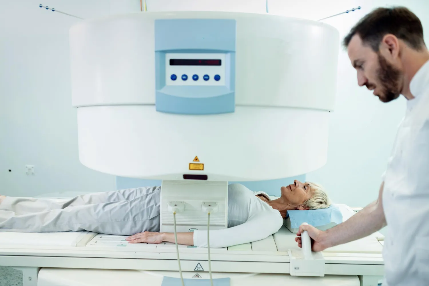
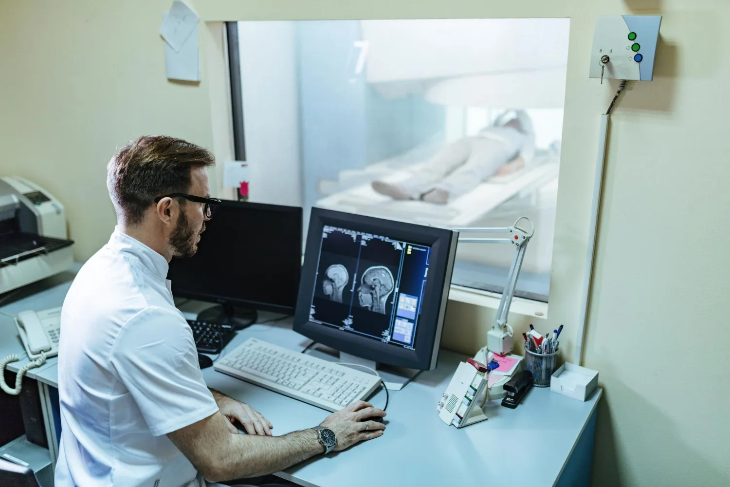
What Happens During the CT Scan?
Many patients feel anxious before undergoing their first CT scan, but the procedure is generally quick, painless, and non-invasive.
Step 1: Registration and Preparation
After confirming your medical details, a radiology technologist explains the procedure and answers any questions you may have.
If contrast material is required, an intravenous cannula is inserted into your arm.
Step 2: Positioning
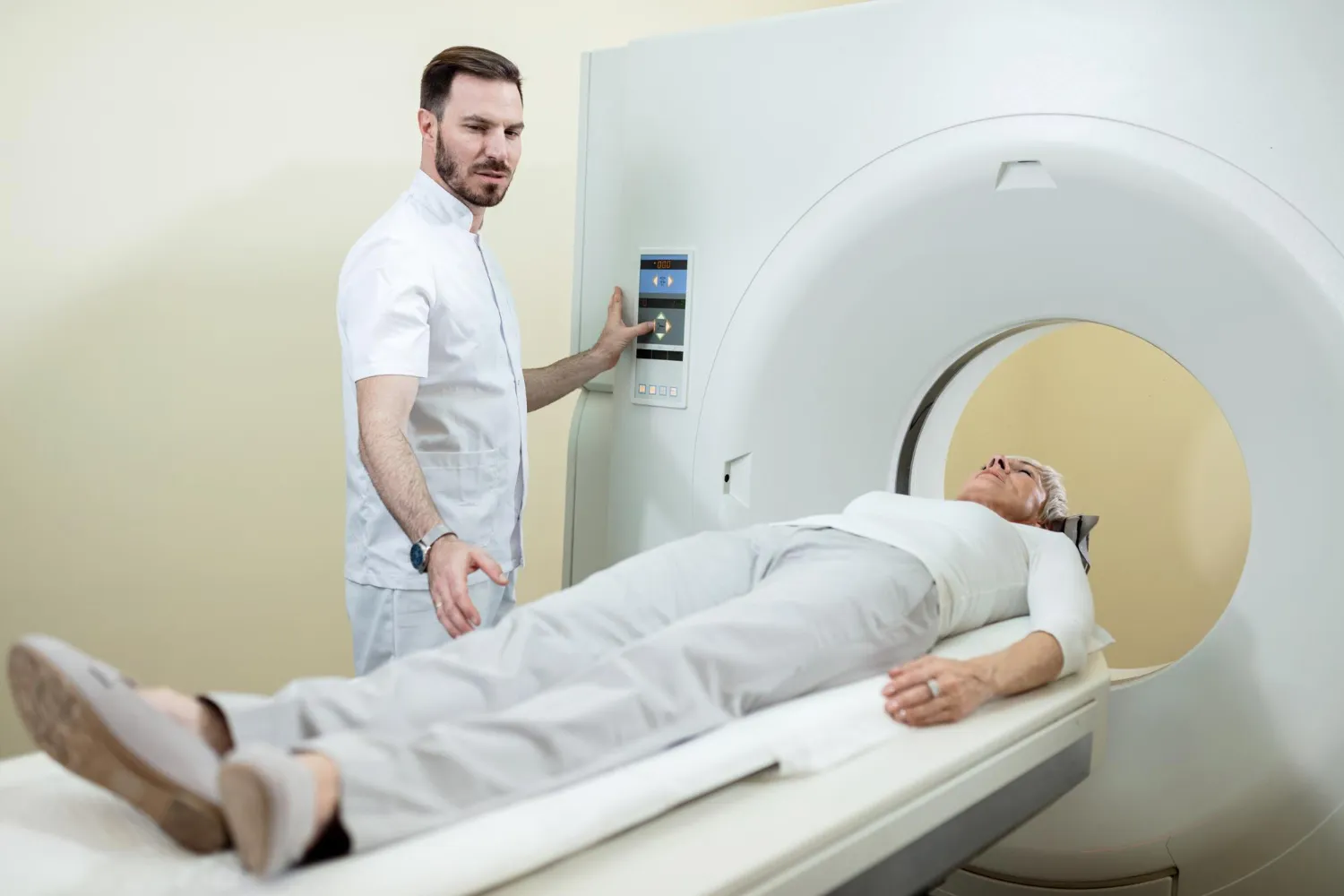
You will lie comfortably on the CT scanning table, usually on your back.
The technologist positions you carefully to ensure the lungs are imaged accurately.
Step 3: Image Acquisition
The table moves slowly through the circular opening of the CT scanner while the X-ray tube rotates around your body.
You may be asked to:
- Hold your breath for a few seconds.
- Remain completely still.
- Follow simple voice instructions.
Remaining still is essential because even slight movement can reduce image clarity.
The actual scan usually takes only a few minutes.
Step 4: After the Scan
Once the examination is complete, you can generally return to your normal daily activities immediately.
Patients who receive contrast dye are often advised to drink extra water to help eliminate the contrast material through the kidneys.
Is a CT Scan Safe?
CT scanning has become one of the safest and most valuable diagnostic tools available in modern medicine.
Like any investigation involving X-rays, a CT scan exposes the body to a controlled amount of ionizing radiation.
However, healthcare providers carefully weigh the small potential risks against the significant benefits of obtaining an accurate diagnosis.
Modern CT scanners use advanced dose-reduction technologies that minimize radiation exposure while maintaining excellent image quality.
For most patients, the benefits of timely diagnosis greatly outweigh the minimal radiation risk.
What Are the Possible Risks?
Complications from CT scanning are uncommon.
However, depending on the type of examination, certain considerations apply.
Radiation Exposure
Although CT scans involve more radiation than standard chest X-rays, the exposure remains within medically accepted safety limits.
Doctors recommend CT imaging only when the expected clinical benefits justify the examination.
Contrast Dye Reactions
Most patients tolerate contrast agents very well.
Occasionally, mild reactions may occur, including:
- Warm sensation during injection
- Metallic taste in the mouth
- Mild nausea
- Temporary flushing
Serious allergic reactions are rare, and radiology departments are fully equipped to manage them promptly.
Kidney Function
Patients with severe kidney disease may require additional evaluation before receiving intravenous contrast.
This is why kidney function tests are sometimes requested before the procedure.
Understanding Your CT Scan Results
Receiving a CT scan report can feel overwhelming, particularly when unfamiliar medical terminology is used.
It is important to remember that the report is interpreted alongside your symptoms, clinical examination, laboratory investigations, and medical history.
A CT scan may identify:
- Lung infections
- Inflammatory changes
- Fibrosis
- Nodules
- Tumours
- Pleural disease
- Blood clots
- Enlarged lymph nodes
- Airway abnormalities
Not every abnormal finding indicates cancer or a serious illness. Many lung abnormalities are benign and may simply require observation or follow-up imaging.
Always discuss your report with your treating physician rather than attempting to interpret it independently.
CT Scan vs Chest X-ray vs MRI
Patients often ask why different imaging tests are recommended for lung conditions.
Each investigation provides different types of information.
| Imaging Test | Primary Use |
|---|---|
| Chest X-ray | Initial evaluation of cough, infection, fractures, and basic lung abnormalities. |
| CT Scan | Detailed assessment of lungs, airways, blood vessels, nodules, tumours, fibrosis, and infections. |
| MRI | Primarily used for evaluating soft tissues, the heart, spine, and certain chest wall abnormalities rather than routine lung imaging. |
Rather than replacing one another, these investigations complement each other depending on the clinical situation.
Can a CT Scan Detect Lung Cancer Early?
Yes. One of the greatest advantages of CT imaging is its ability to detect very small lung nodules that may not be visible on a conventional chest X-ray.
In selected high-risk individuals—particularly long-term smokers—Low-Dose CT (LDCT) has become an important tool for lung cancer screening because it can identify cancer at an earlier and more treatable stage.
However, not every lung nodule represents cancer. Many nodules result from previous infections, inflammation, or benign conditions.
Further evaluation depends on factors such as:
- Nodule size
- Shape
- Growth over time
- Patient age
- Smoking history
- Family history
Your physician will determine whether additional imaging, biopsy, or routine follow-up is appropriate.
Tips for Maintaining Healthy Lungs
Although some lung diseases cannot be prevented, adopting healthy habits can significantly reduce the risk of respiratory illness.
Stop Smoking
Smoking remains the leading preventable cause of lung disease and lung cancer.
Quitting smoking provides immediate and long-term health benefits.
Avoid Air Pollution
Whenever possible:
- Wear protective masks in polluted environments.
- Improve indoor ventilation.
- Reduce exposure to occupational dust and chemicals.
Stay Physically Active
Regular exercise strengthens the lungs, improves breathing efficiency, and enhances overall cardiovascular fitness.
Keep Vaccinations Updated
Influenza and pneumococcal vaccinations reduce the risk of serious respiratory infections, particularly in older adults and individuals with chronic lung disease.
Seek Medical Attention Early
Persistent cough, unexplained weight loss, coughing up blood, prolonged fever, or worsening breathlessness should never be ignored.
Early medical evaluation often leads to earlier diagnosis and more effective treatment.
Conclusion
A CT scan has transformed the way lung diseases are diagnosed by allowing doctors to detect abnormalities with remarkable precision. From persistent cough and recurrent infections to pulmonary fibrosis, lung nodules, and suspected lung cancer, CT imaging provides detailed information that often cannot be obtained through a conventional chest X-ray alone.
Early diagnosis is essential for effective treatment, and advanced imaging plays a key role in identifying disease before complications develop. If your doctor recommends a CT scan, understanding the purpose of the examination can help you approach the procedure with confidence.
At Truscan Diagnostics, we are dedicated to delivering advanced diagnostic imaging supported by experienced radiologists, modern technology, and patient-centred care. Our commitment to quality ensures that every scan contributes to accurate diagnosis and better healthcare outcomes.
Schedule your CT scan at Truscan Diagnostics and take an informed step towards better respiratory health.
Medical Disclaimer
This article is intended for educational purposes only and should not replace professional medical advice, diagnosis, or treatment. Always consult your treating physician regarding your symptoms, imaging results, or healthcare decisions.
Frequently Asked Questions
Everything you need to know about our service
How long does a CT scan of the lungs take?
The actual scan usually takes only a few minutes, although the complete appointment may last between 15 and 30 minutes depending on preparation and whether contrast is required.
Is a CT scan painful?
No. A CT scan is completely painless. If contrast dye is administered, you may briefly experience a warm sensation or metallic taste.
Can I eat before the scan?
For a plain CT scan, eating is generally allowed. If contrast is required, your doctor or imaging centre may recommend fasting for a few hours.
Will I receive my report immediately?
Report preparation depends on the complexity of the examination. Your radiologist will carefully analyse the images before issuing the final report.
Does every lung nodule indicate cancer?
No. Many lung nodules are benign. Your doctor will interpret the findings based on your medical history, symptoms, and, if required, follow-up imaging.
Can children undergo CT scans?
Yes, but only when medically necessary. Radiation doses are carefully adjusted for children, and alternative imaging methods may be considered whenever appropriate.
Why Choose Truscan Diagnostics?
Accurate imaging plays a vital role in diagnosing and managing lung diseases. At Truscan Diagnostics, we combine advanced CT imaging technology with experienced radiologists to provide detailed, high-quality diagnostic evaluations that support timely and informed clinical decisions.
Our imaging facilities are designed to prioritise patient comfort, safety, and precision. Whether you require a routine chest CT, High-Resolution CT (HRCT), contrast-enhanced CT, or specialised pulmonary imaging, our team follows established quality protocols to deliver reliable results with minimal waiting time.
From appointment scheduling to report delivery, we are committed to providing a seamless diagnostic experience backed by compassionate patient care.