
Beyond the Scan: How Non-Invasive Imaging Helps Detect Health Problems Early
Non-invasive imaging techniques allow healthcare professionals to examine organs, tissues, bones and other structures without conventional surgery. Depending on the clinical question, imaging may include X-ray, ultrasound, CT scan or MRI.
Each imaging method works differently and provides different types of information. Choosing the right scan is therefore not simply about selecting the most advanced technology. It is about choosing an examination that is appropriate for the person’s symptoms, medical history and clinical needs.
What Is Non-Invasive Medical Imaging?
Non-invasive medical imaging refers to techniques used to produce images or information about structures inside the body without requiring conventional surgical access.
Medical imaging can help healthcare professionals:
- Investigate symptoms
- Identify structural abnormalities
- Evaluate injuries
- Support diagnosis
- Monitor certain medical conditions
- Assess treatment response
- Plan some medical procedures
The FDA describes medical imaging as a group of technologies used to view the body to help diagnose, monitor or treat medical conditions. Different imaging technologies provide different information about the area being examined.
Some imaging methods use ionizing radiation, while others do not.
Why Is Medical Imaging Important for Early Detection?
Some conditions may not produce obvious symptoms at an early stage, while others may cause symptoms that overlap with many different conditions.
Imaging can provide additional information that may help a healthcare professional investigate a clinical concern.
For example, depending on the situation:
- An X-ray may help evaluate bones or the chest.
- An ultrasound may examine organs, soft tissues or blood flow.
- A CT scan can produce detailed cross-sectional images.
- An MRI can provide detailed images of soft tissues and other structures.
Early imaging does not necessarily mean that every disease will be detected before symptoms appear. Imaging is most useful when it is appropriately selected for a specific clinical question.
What Are the Main Types of Non-Invasive Imaging?
1. X-Ray
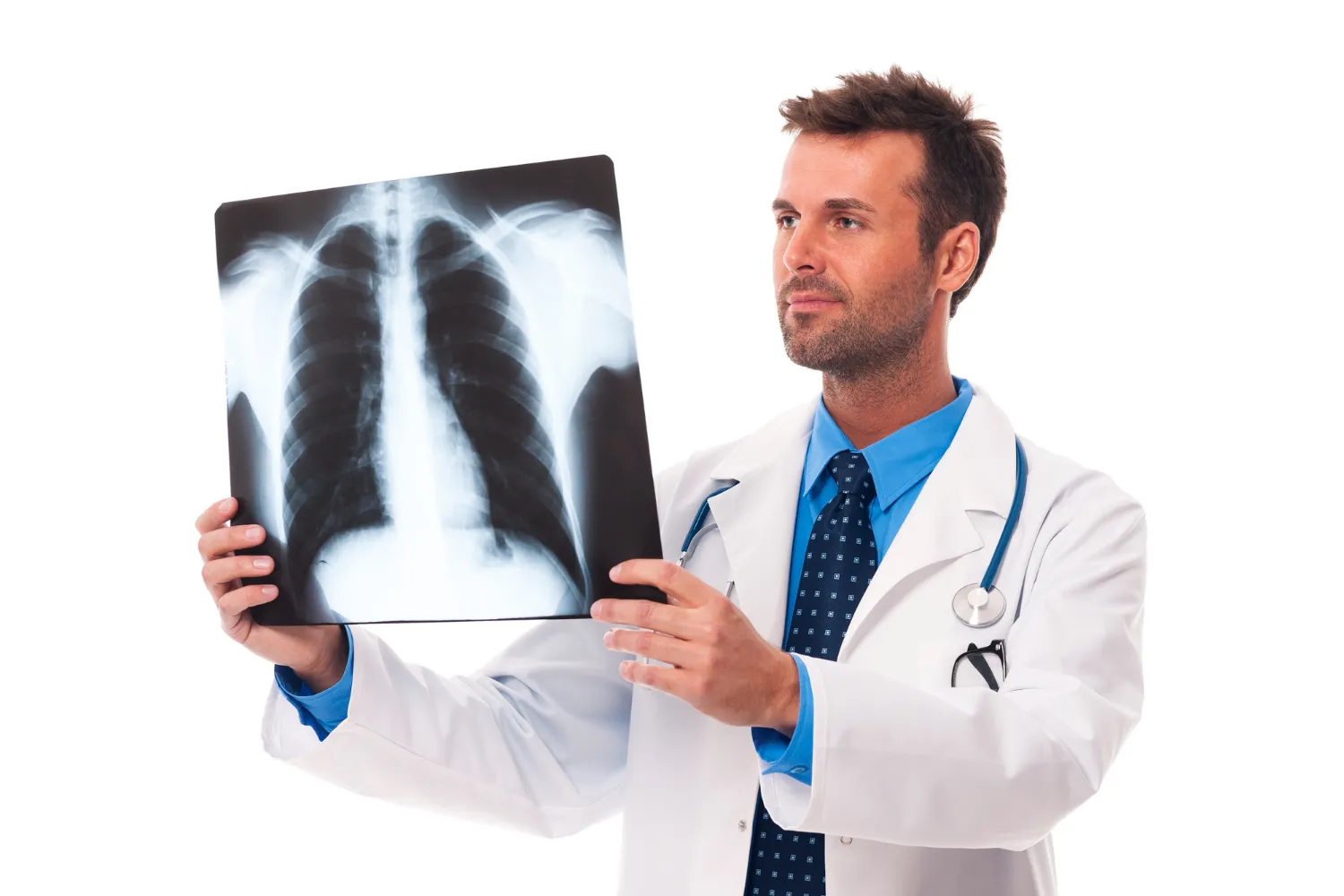
An X-ray is one of the most commonly used medical imaging techniques.
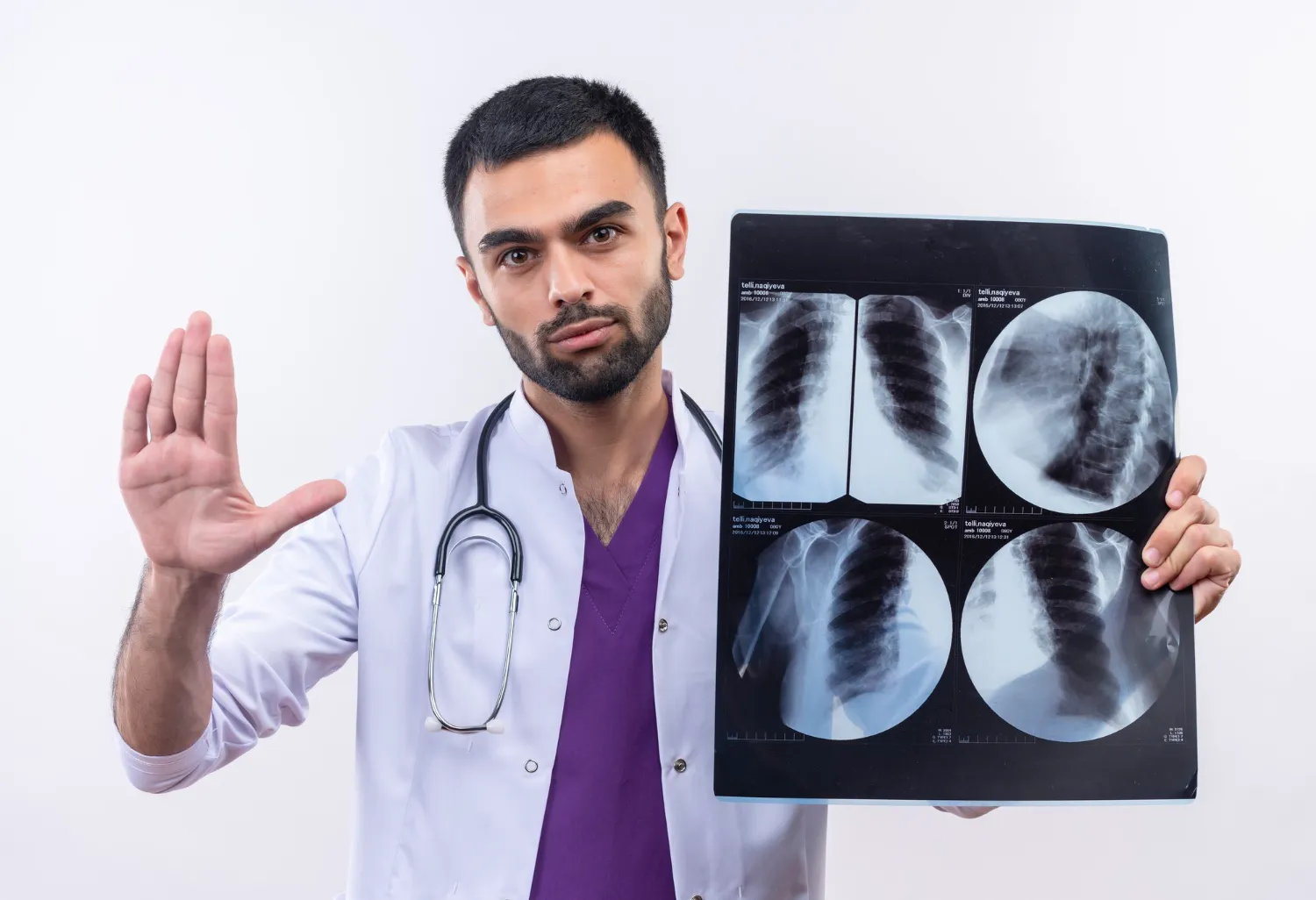
It uses X-rays to produce images of structures inside the body. X-rays are particularly useful for evaluating bones and can also be used for areas such as the chest.
Common uses may include evaluating:
- Bone injuries and fractures
- Chest conditions
- Certain joint problems
- Some structural abnormalities
A conventional X-ray generally produces a two-dimensional image.
Because X-rays use ionizing radiation, the examination should be performed when medically appropriate. The amount of radiation varies according to the type of examination.
2. Ultrasound
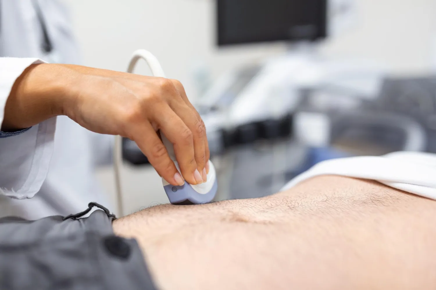
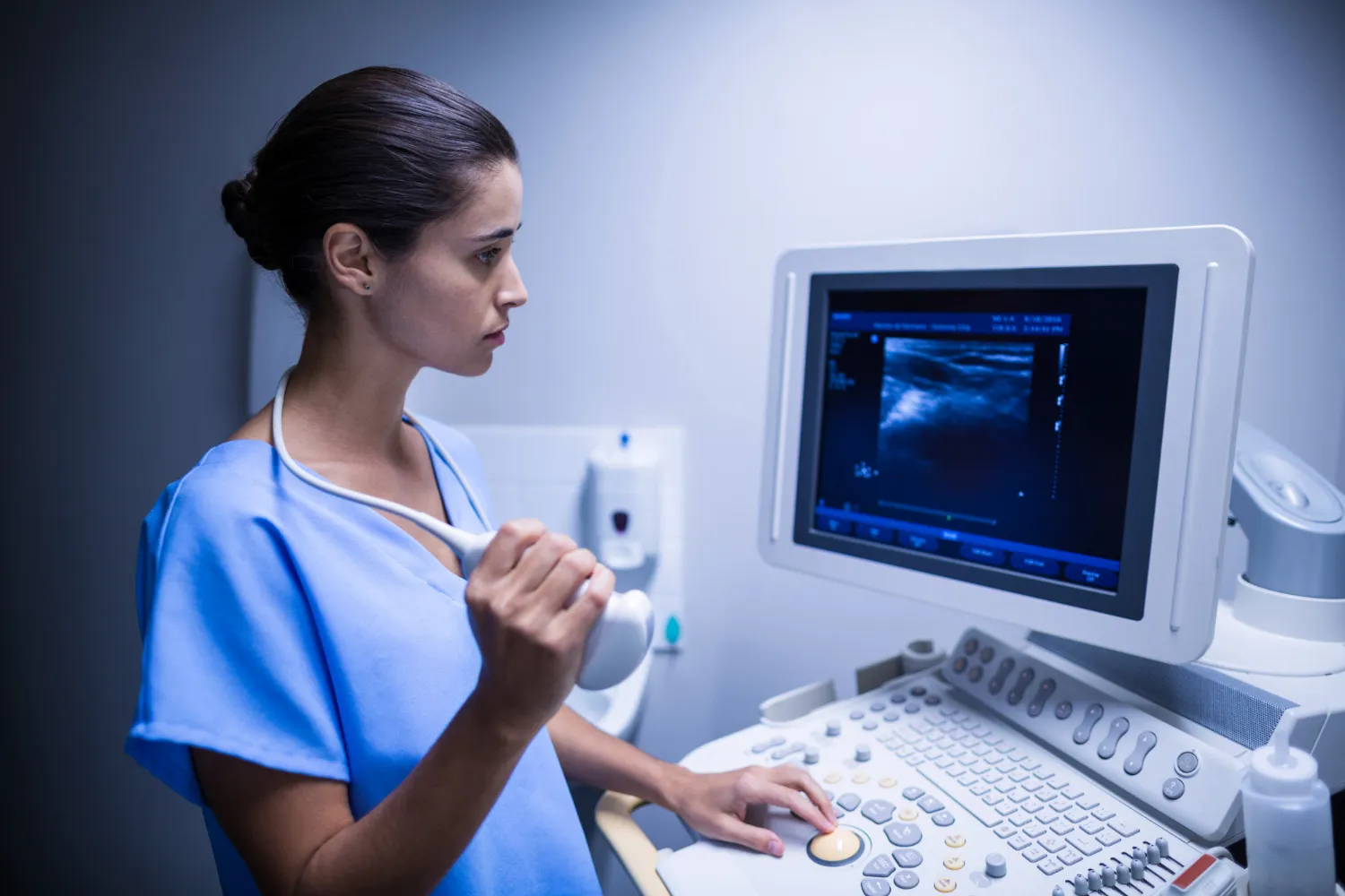
Ultrasound imaging, also known as sonography, uses high-frequency sound waves to produce images of structures inside the body.
Unlike X-rays and CT scans, ultrasound does not use ionizing radiation.
Ultrasound can be used to examine areas such as:
- Abdominal organs
- Soft tissues
- Blood vessels
- The thyroid
- Certain musculoskeletal structures
- Reproductive organs
- The heart through echocardiography
Doppler ultrasound can also provide information about blood flow.
One advantage of ultrasound is that images can be captured in real time, allowing movement and blood flow to be assessed in appropriate examinations.
3. CT Scan
A CT scan, or computed tomography scan, uses X-rays and computer processing to create cross-sectional images of the body.
Unlike a conventional X-ray, which generally produces a projection image, CT produces multiple cross-sectional images or “slices” that can provide more detailed information about internal structures.
CT can be used to evaluate many areas of the body and may help healthcare professionals investigate:
- Injuries and trauma
- Internal bleeding
- Certain abdominal conditions
- Chest conditions
- Bone abnormalities
- Some cancers
- Blood vessels and other structures
CT can also be useful for treatment planning and monitoring certain conditions.
However, CT uses ionizing radiation. In some examinations, a contrast agent may also be used to improve visualization. The decision to perform a CT should consider the expected medical benefit and potential risks.
4. MRI Scan
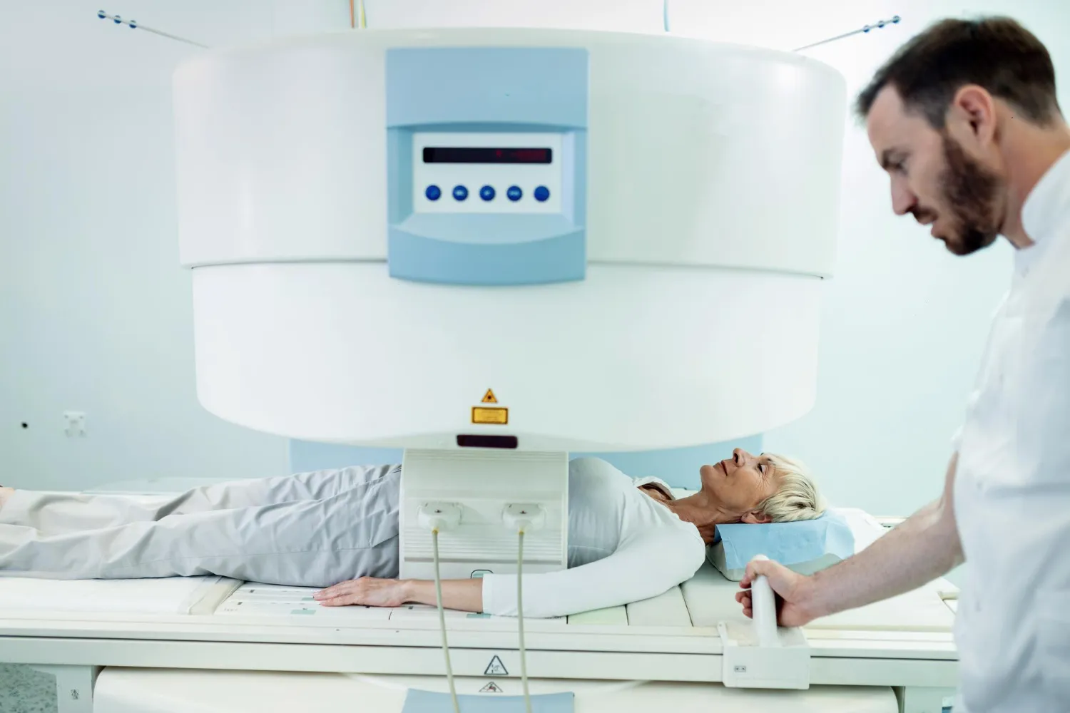
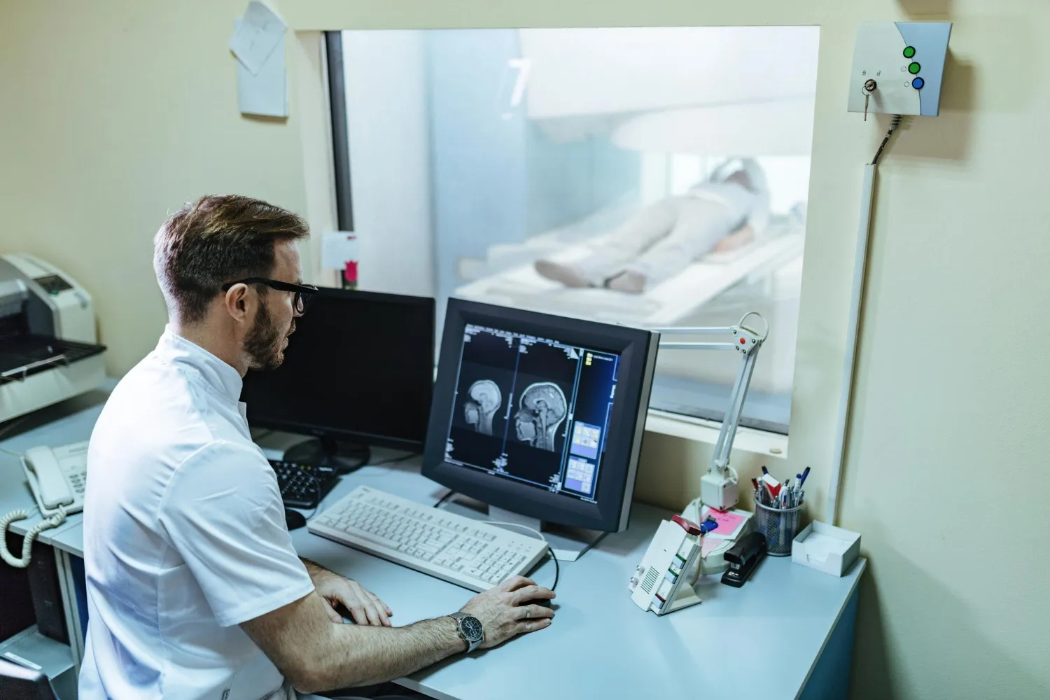
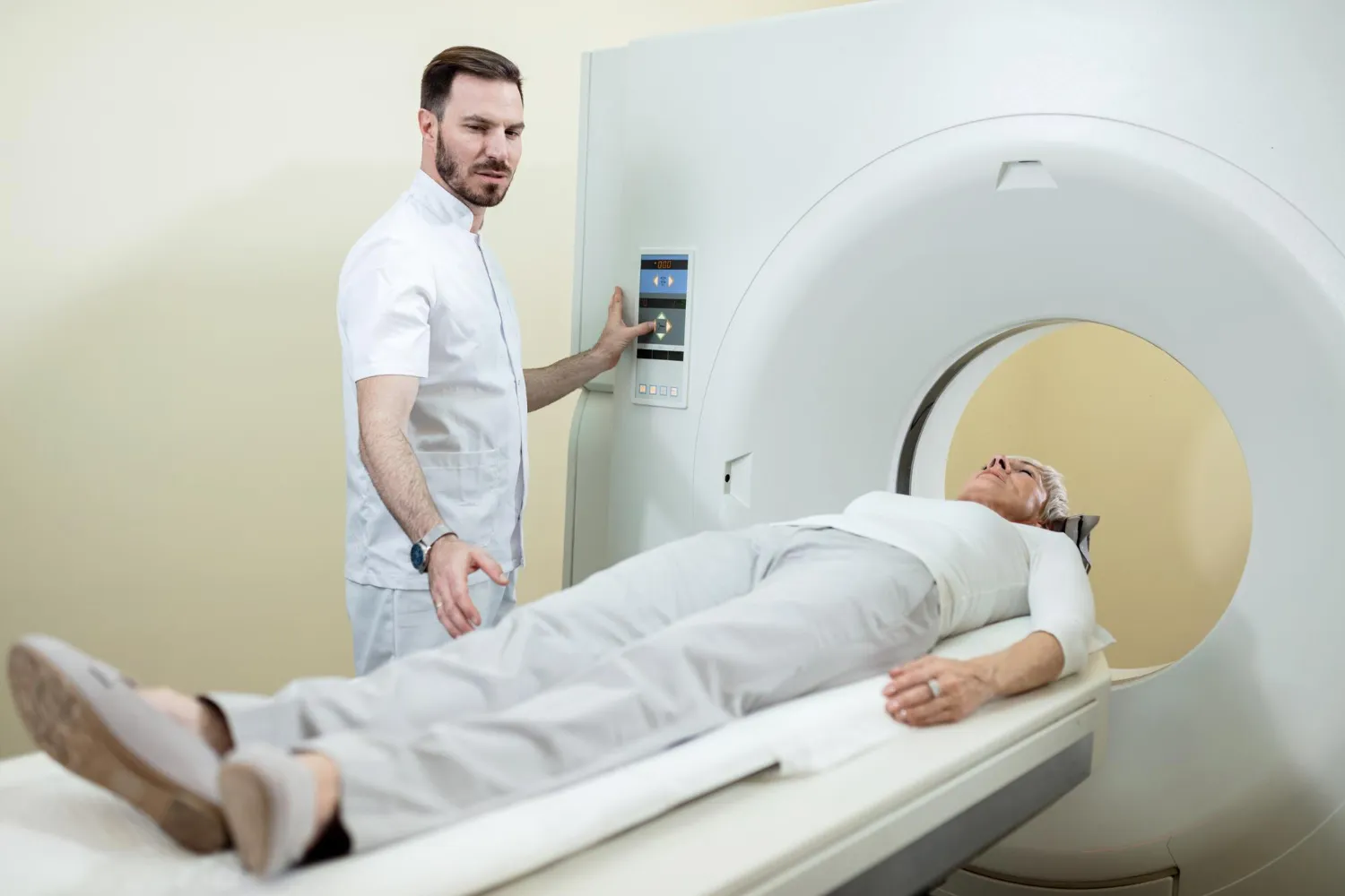
Magnetic Resonance Imaging (MRI) uses strong magnetic fields and radiofrequency energy to create detailed images of structures inside the body.
MRI does not use ionizing radiation. It is particularly useful for producing detailed images of many soft tissues, although its suitability depends on the body part and clinical question.
MRI may be used to evaluate areas such as:
- Brain and spinal cord
- Joints and muscles
- Ligaments and tendons
- Certain abdominal and pelvic structures
- Blood vessels
- Other soft tissues
MRI examinations can take longer than some other imaging tests, and patients with certain implants or metal objects may require special safety assessment before the examination.
X-Ray vs Ultrasound vs CT vs MRI
Each imaging method has a different role.
| Imaging Test | How It Works | Common Strength |
|---|---|---|
| X-Ray | Uses X-rays | Bones and chest imaging |
| Ultrasound | Uses sound waves | Soft tissues, organs and blood flow |
| CT Scan | Uses X-rays and computer reconstruction | Detailed cross-sectional imaging |
| MRI | Uses magnetic fields and radiofrequency energy | Detailed soft-tissue imaging |
The “best” scan depends on what the healthcare professional needs to evaluate.
A more advanced scan is not automatically a more appropriate scan.
Can Imaging Detect Health Problems Before Symptoms Become Severe?
Imaging can sometimes identify abnormalities that require further evaluation, but its ability to detect a condition depends on the disease, the imaging method and the reason for performing the examination.
For people with symptoms, imaging can help investigate a suspected condition and provide information that may guide further management.
For people without symptoms, however, not every imaging test is appropriate as a general screening examination.
For example, the FDA states that it does not currently see a demonstrated benefit for whole-body CT scanning of individuals without symptoms and highlights the potential for radiation exposure and unnecessary follow-up from incidental findings.
This is why screening decisions should be based on established medical recommendations and individual risk rather than simply choosing a scan for reassurance.
What Health Problems Can Medical Imaging Help Investigate?
Depending on the imaging method and clinical situation, medical imaging may help investigate:
Bone and Joint Problems
X-ray, CT and MRI can provide different types of information about bones, joints and surrounding tissues.
For example, X-ray may be used to evaluate fractures, while MRI can provide more detailed information about soft tissues such as ligaments and tendons.
Brain and Neurological Conditions
CT and MRI can provide images of the brain and other neurological structures.
Depending on the situation, imaging may be used to investigate symptoms such as severe headaches, neurological changes, suspected stroke, trauma or other concerns.
The appropriate scan depends on the urgency and clinical question.
Abdominal and Pelvic Conditions
Ultrasound, CT and MRI may be used to examine abdominal and pelvic organs.
For example, ultrasound can be used to assess certain organs and structures, while CT or MRI may provide additional information when more detailed imaging is required.
Chest and Lung Conditions
Chest X-rays and CT scans can provide information about the lungs and other structures in the chest.
A healthcare professional may recommend a particular examination based on symptoms, medical history and suspected conditions.
Heart and Blood Vessels
Certain imaging techniques can also be used to evaluate cardiovascular structures.
Ultrasound-based echocardiography can provide information about the heart’s structure and movement, while other imaging techniques may be used to evaluate blood vessels or specific cardiac conditions when clinically indicated.
Is Medical Imaging Always Safe?
Medical imaging is widely used, but each technique has its own benefits, limitations and safety considerations.
X-Ray and CT
X-ray-based imaging uses ionizing radiation.
The FDA recommends that healthcare professionals ensure that X-ray examinations are medically justified and consider alternatives that use less or no ionizing radiation, such as ultrasound or MRI, when medically appropriate.
CT generally provides substantial diagnostic benefits when appropriately used, but it involves more radiation than a conventional X-ray.
Ultrasound
Ultrasound does not use ionizing radiation and is widely used for diagnostic purposes. However, it should still be used appropriately by trained healthcare professionals.
MRI
MRI does not use ionizing radiation, but its strong magnetic field creates specific safety considerations.
Patients should inform the healthcare team about implants, metal objects, medical devices or other relevant conditions before an MRI examination.
How Should You Prepare for an Imaging Test?
Preparation depends on the type of examination.
Some imaging tests require little or no preparation, while others may involve instructions such as:
- Fasting before certain examinations
- Drinking water or having a full bladder for selected ultrasound examinations
- Removing metal objects before MRI
- Informing the imaging team about implants or medical devices
- Following specific instructions if contrast material is planned
Patients should follow the preparation instructions provided by their healthcare provider or diagnostic centre.
Do not assume that preparation requirements are the same for every CT, MRI or ultrasound examination.
What Happens After the Scan?
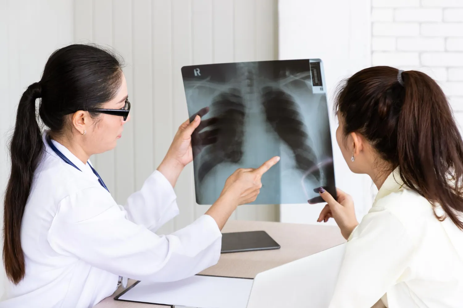
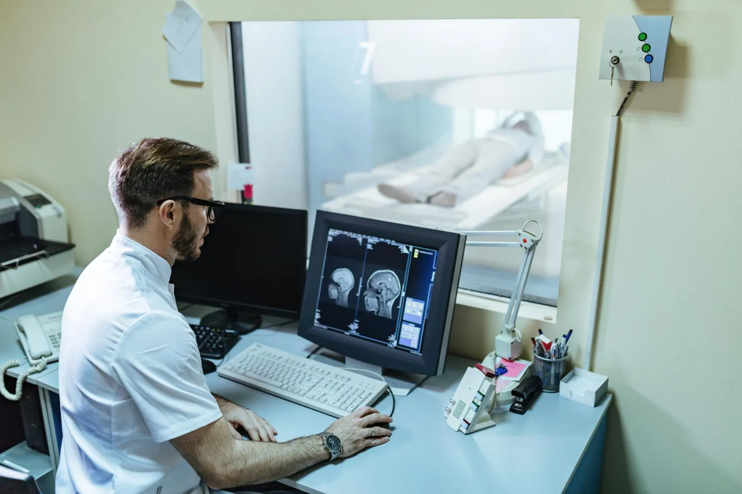
After an imaging examination, the images are reviewed and interpreted by an appropriately qualified healthcare professional.
The imaging report is considered together with:
- Symptoms
- Medical history
- Physical examination
- Laboratory results
- Previous imaging
- Other diagnostic findings
An imaging result should not be interpreted in isolation.
A finding on a scan may require additional testing, monitoring or clinical follow-up. In some cases, an imaging result may be normal even when symptoms require further evaluation.
Can a Normal Scan Guarantee That Everything Is Fine?
No.
Medical imaging is an important diagnostic tool, but no imaging test can detect every possible health condition.
Different tests have different strengths and limitations. Some conditions may not be visible on a particular scan, while some findings may require additional investigation.
A normal imaging report should therefore be understood in the context of the reason the scan was performed and the person’s overall clinical picture.
If symptoms continue despite a normal scan, further medical evaluation may still be appropriate.
Why Choosing the Right Imaging Test Matters
More imaging does not necessarily mean better healthcare.
The appropriate test should answer a specific clinical question while considering factors such as:
- Symptoms
- Age
- Medical history
- Pregnancy status when relevant
- Previous imaging
- Suspected condition
- Radiation exposure
- Whether contrast is required
- The information needed by the healthcare professional
For example, if a question can be appropriately answered with an imaging method that does not use ionizing radiation, that option may sometimes be considered. The FDA specifically recommends considering alternatives such as ultrasound or MRI when medically appropriate for reducing unnecessary radiation exposure.
Conclusion
Non-invasive medical imaging has become an important part of modern healthcare. X-ray, ultrasound, CT and MRI each provide different types of information and can help healthcare professionals investigate symptoms, evaluate injuries, identify abnormalities and monitor selected conditions.
However, imaging should not be viewed as a replacement for clinical evaluation or as a guarantee that every health problem will be detected.
The most useful scan is the one that appropriately answers the clinical question while considering the benefits, limitations and safety factors of the examination.
If you have persistent or concerning symptoms, speak with a qualified healthcare professional about whether imaging is appropriate and which examination may provide the information needed.
TruScan Diagnostics provides diagnostic imaging services that can support healthcare professionals in evaluating a range of health concerns. The appropriate imaging test should be selected according to individual clinical needs and interpreted in the context of the patient’s overall health.
Related Reading: MRI vs CT Scan | MRI, CT Scan, or Ultrasound | What is a CT Scan? | Cardiac Diagnostic Tests
Book an Imaging Test
TruScan Diagnostics offers X-ray, ultrasound, CT and MRI at our JP Nagar centres, open 24 hours. Call +91 8956 600 600 to book.
Medical Disclaimer: This article is intended for educational purposes only and should not replace professional medical advice, diagnosis, or treatment. Always consult your treating physician regarding your symptoms, test results, or healthcare decisions.
TruScan Diagnostics, JP Nagar, Bengaluru
- JP Nagar 7th Phase: Manvi Arcade, WGHCS Layout, behind Ever Fine Supermarket, JP Nagar 7th Phase, Bengaluru 560078
- JP Nagar 2nd Phase: 213, 9th Cross Rd, KSRTC Layout, JP Nagar 2nd Phase, Bengaluru 560078
- Phone: +91 8956 600 600 (WhatsApp available)
- Website: truscan.in
- Instagram: @truscandiagnosticsofficial
- Facebook: facebook.com/truscandiagno
- Open 24 Hours
Frequently Asked Questions About Non-Invasive Imaging
Useful answers about X-ray, ultrasound, CT and MRI
What is non-invasive medical imaging?
Non-invasive medical imaging uses technologies such as X-ray, ultrasound, CT and MRI to obtain information about structures inside the body without conventional surgery.
Which imaging test is best?
There is no single best imaging test for every condition. The appropriate examination depends on the symptoms, suspected condition and information required.
Is an MRI safer than a CT scan?
MRI does not use ionizing radiation, whereas CT uses X-rays and therefore involves ionizing radiation. However, MRI has its own safety considerations, particularly related to its strong magnetic field. The appropriate examination depends on the clinical situation.
Does ultrasound use radiation?
Ultrasound uses high-frequency sound waves rather than ionizing radiation.
Does a CT scan detect all diseases?
No. CT can provide detailed images and is useful for many diagnostic purposes, but it cannot detect every disease or condition.
Can medical imaging detect disease before symptoms appear?
Some imaging examinations can identify abnormalities before symptoms become severe, but imaging is not a universal screening tool. Screening should be performed when there is evidence that the particular test provides meaningful benefit for the relevant population.
Is a full-body CT scan recommended for everyone?
No. The FDA states that it has not found evidence that whole-body CT screening of asymptomatic individuals provides more benefit than harm.
How often should I get an imaging test?
There is no universal frequency for all imaging tests. The need and timing depend on the individual's health, symptoms, risk factors, previous results and medical recommendations.