

How Radiography Helps Detect Problems Early: Understanding X-ray, CT and MRI Scans

Many health conditions develop gradually, and some may not produce noticeable symptoms during their early stages. When a person experiences persistent pain, unexplained discomfort, an injury or other concerning symptoms, medical imaging can provide valuable information about what is happening inside the body.
Radiography is one of the most widely used diagnostic imaging techniques. It uses X-rays to create images of internal structures, helping doctors examine bones, joints, the chest and certain other body regions.
Modern diagnostic imaging has expanded beyond conventional X-rays. Technologies such as Computed Tomography (CT), Magnetic Resonance Imaging (MRI) and ultrasound provide different types of information about organs, tissues and internal structures.
With appropriate clinical assessment, these investigations can support early diagnosis, identify abnormalities and help doctors plan further evaluation or treatment.
Understanding how radiography helps in early detection can help patients make informed decisions about diagnostic investigations and recognise why choosing the right imaging test matters.
1. What Is Diagnostic Radiography?
Diagnostic radiography is a medical imaging technique that uses X-rays to produce images of structures inside the body.
X-rays pass through the body and are absorbed differently by different tissues.
For example:
- Bones absorb more X-rays and generally appear white.
- Soft tissues appear in different shades of grey.
- Air-filled areas appear darker.
These differences allow radiologists to examine internal structures and identify certain abnormalities.
Traditional film-based X-rays have largely been replaced by digital radiography in many healthcare settings.
What Is Digital Radiography?
Digital radiography uses electronic detectors to capture X-ray images.
The images can be viewed on computer systems, stored digitally and shared with authorised healthcare professionals.
Potential advantages include:
- Faster image availability.
- Digital image storage.
- Convenient image retrieval.
- Easier sharing for medical review.
- Opportunities for image processing and dose optimisation.
The diagnostic quality still depends on appropriate positioning, imaging protocols, equipment and professional interpretation.
2. How Does Radiography Help with Early Detection?
Medical imaging contributes to diagnosis by providing visual information that may not be available through physical examination alone.
Its usefulness depends on the suspected condition and the type of imaging selected.
Detecting bone and joint abnormalities
X-rays can help identify fractures, joint alignment problems, certain degenerative changes and other bone abnormalities.
For example, a person experiencing persistent joint pain may require imaging to investigate structural changes.
Early evaluation can help the doctor decide whether additional assessment, treatment or specialist referral is needed.
Evaluating lung and chest conditions
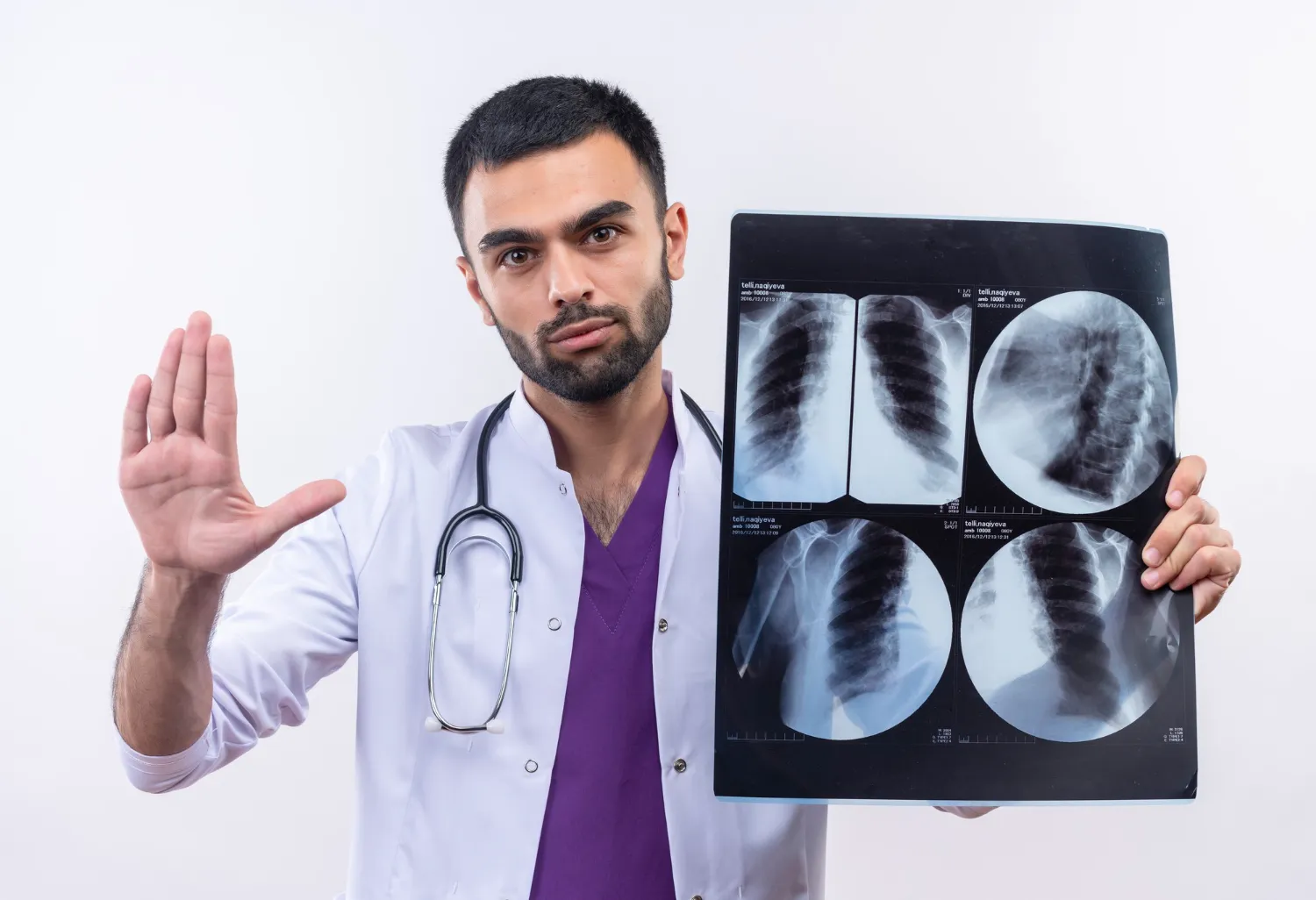
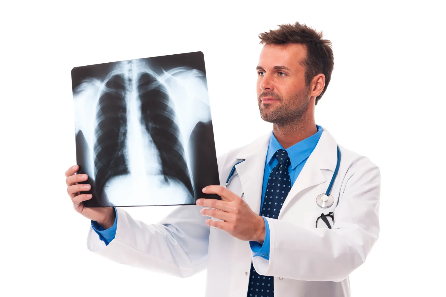
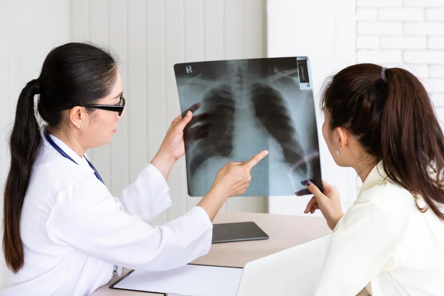
Chest X-rays help doctors investigate symptoms such as persistent cough, fever, chest pain and breathing difficulty.
They may reveal abnormalities associated with pneumonia, fluid around the lungs and certain other chest conditions.
Investigating unexplained symptoms
When symptoms cannot be fully explained through physical examination, imaging may provide additional diagnostic information.
For example, persistent abdominal pain may require ultrasound or CT depending on the suspected cause.
Supporting treatment planning
Imaging can help doctors understand the location and extent of certain abnormalities.
It may also be used to monitor selected conditions and evaluate changes over time.
However, medical imaging is not a replacement for clinical examination or laboratory testing.
3. Different Types of Medical Imaging for Early Diagnosis
Different imaging techniques are designed to answer different clinical questions.
Choosing the appropriate test is important because no single imaging method is suitable for every condition. A closer look at MRI and CT, and at how non-invasive imaging is chosen, can help explain why the test depends on the question the doctor needs to answer.
A. Digital X-ray
Digital X-ray is commonly used to examine bones, joints and the chest.
It may help investigate:
- Bone fractures.
- Joint abnormalities.
- Certain lung infections.
- Changes in bone structure.
- Some chest abnormalities.
For example, following an injury, a doctor may recommend an X-ray to check for a fracture.
A chest X-ray may be recommended for selected patients with persistent cough, fever or suspected lung infection.
However, X-rays cannot identify every bone, lung or soft-tissue condition. Additional investigations may be required when symptoms persist despite normal findings.
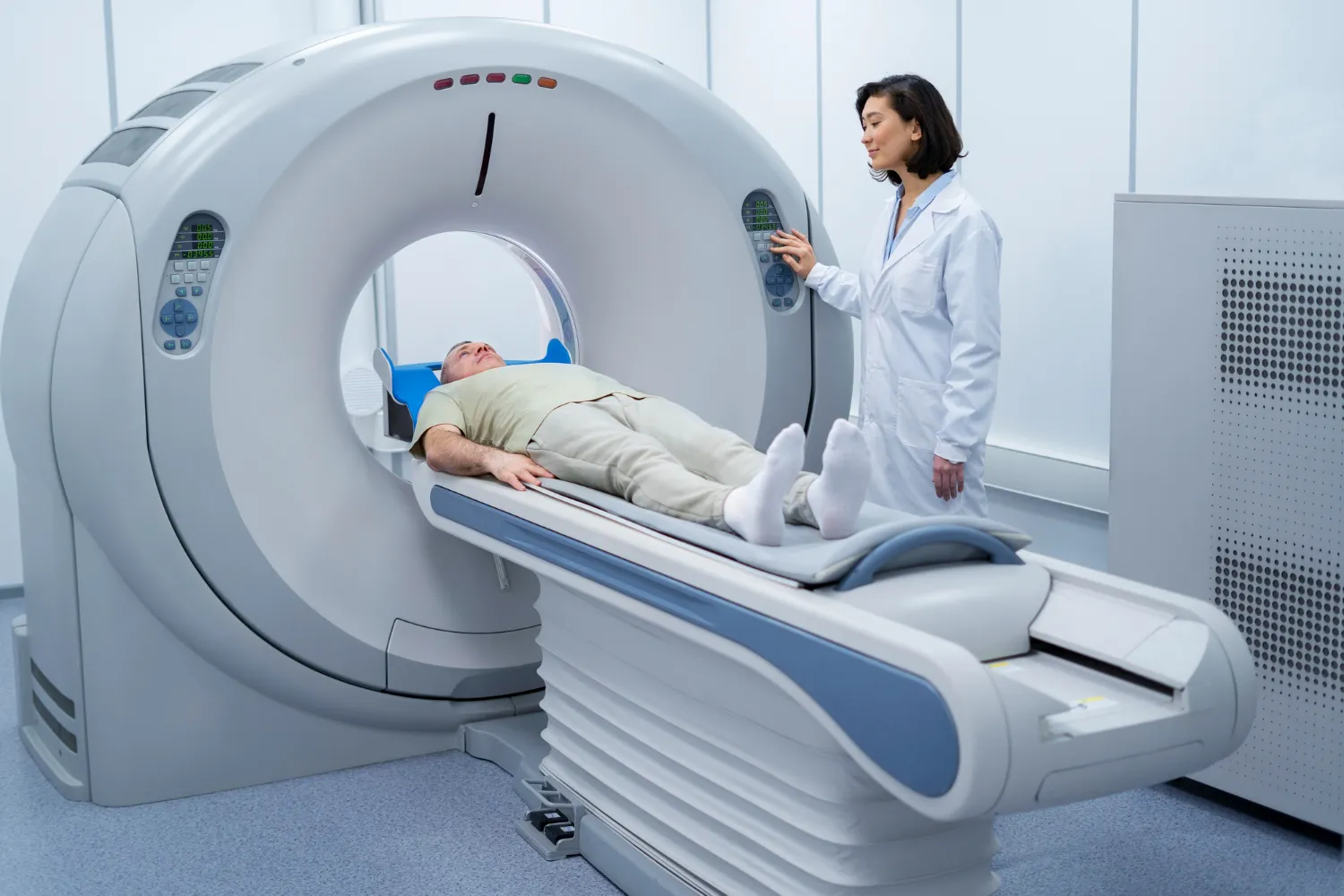
B. CT Scan (Computed Tomography)
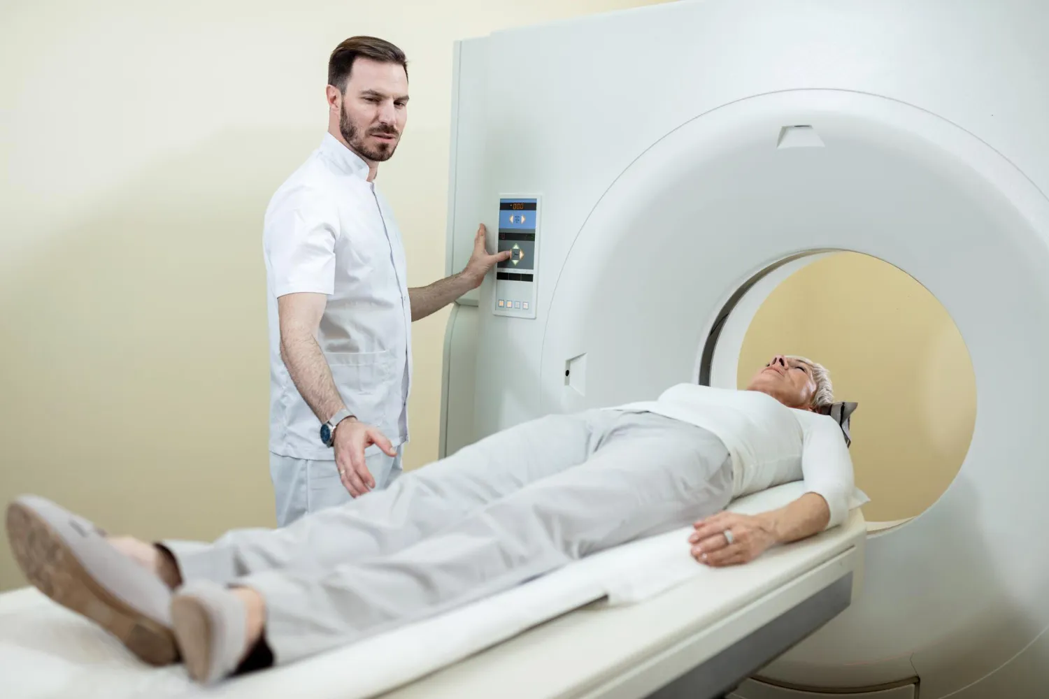
A CT scan uses X-rays and computer processing to create detailed cross-sectional images of the body. What a CT scan involves depends on the body region and the clinical question.
Unlike conventional X-rays, which produce projection images, CT scans provide multiple image sections that can be reconstructed into different views.
Doctors may recommend CT imaging to investigate:
- Internal injuries.
- Certain lung abnormalities. A CT scan for lung disorders may be considered when a chest X-ray does not provide enough detail.
- Abdominal conditions.
- Complex fractures.
- Certain suspected tumours.
- Selected neurological emergencies.
CT imaging can provide valuable information when more detailed assessment is needed.
However, CT scans involve ionising radiation. The decision to perform one should be based on clinical need.
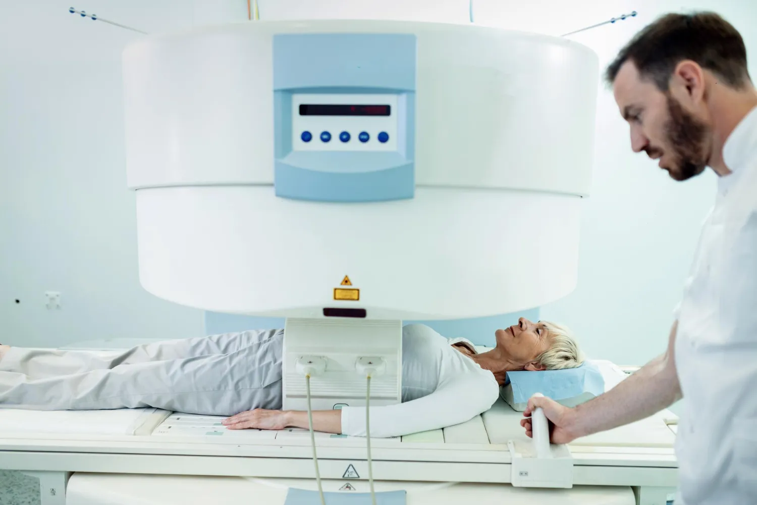
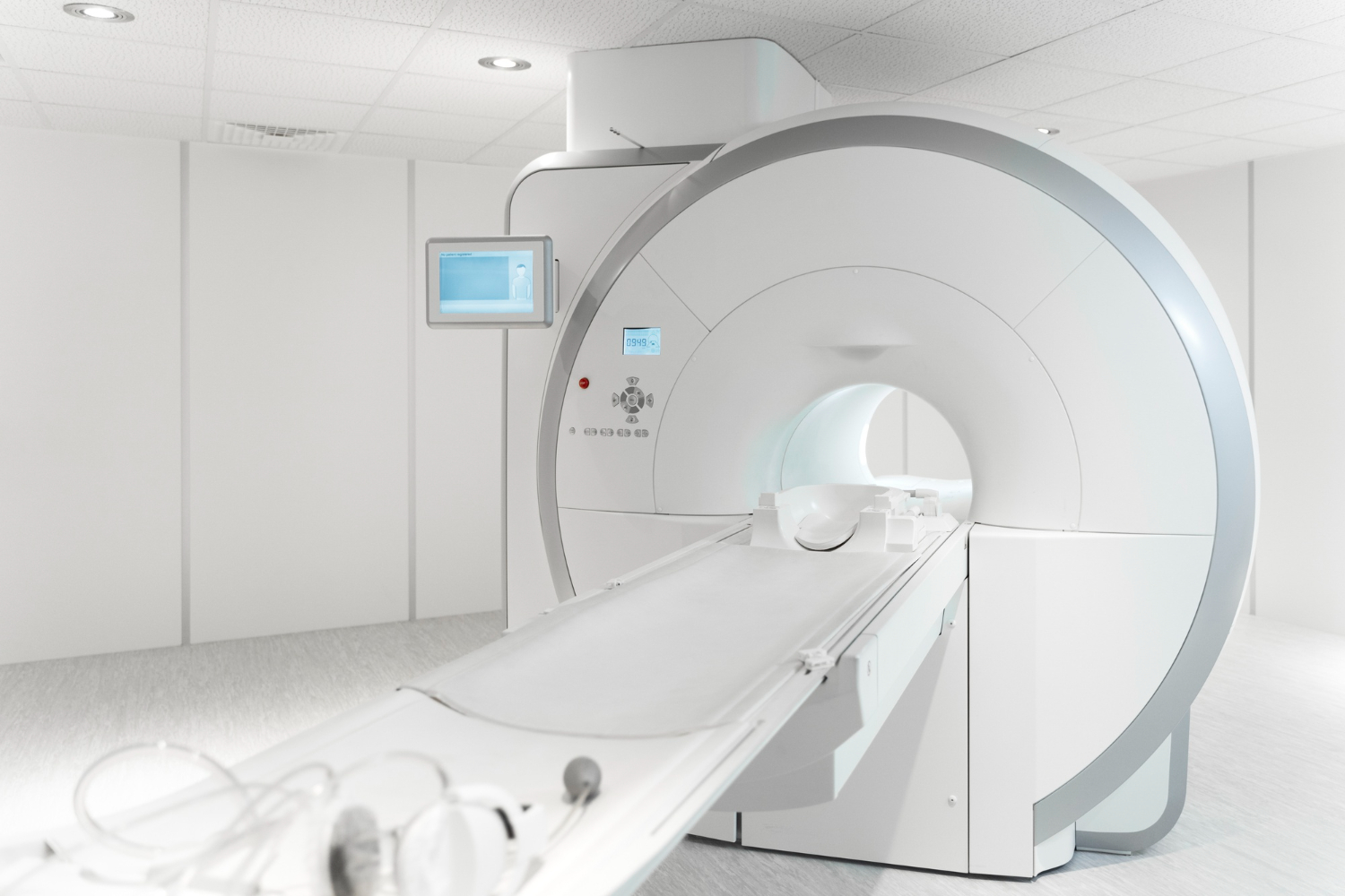
C. MRI Scan (Magnetic Resonance Imaging)
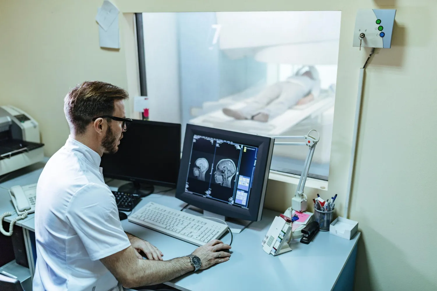
MRI uses strong magnetic fields and radiofrequency waves to produce detailed images of internal structures.
It does not use ionising radiation.
MRI is particularly useful for evaluating:
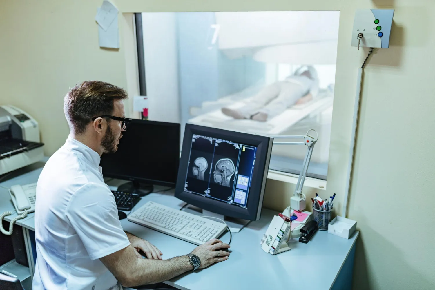
- Brain and spinal structures.
- Muscles and ligaments.
- Joints and soft tissues.
- Certain internal organs.
- Selected neurological conditions.
For example, an MRI may be recommended when a doctor needs detailed information about spinal structures or certain joint injuries.
Some patients cannot undergo certain MRI examinations because of specific implanted devices or other safety considerations. The radiology team must be informed about relevant medical history.
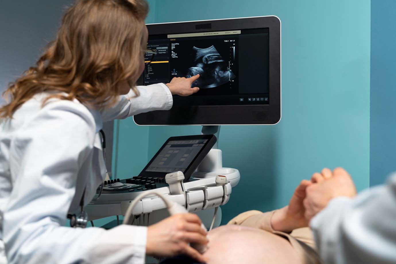
D. Ultrasound Scan
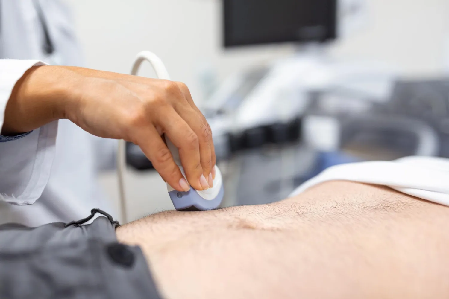
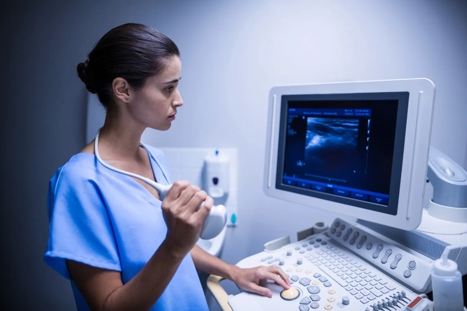
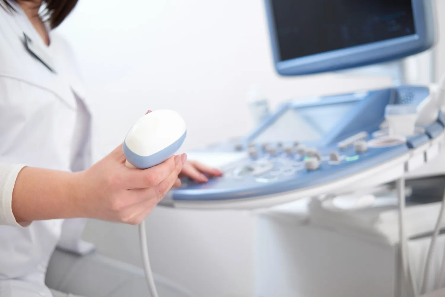
Ultrasound uses high-frequency sound waves to produce images of internal structures.
It is commonly used for:
- Abdominal imaging.
- Pelvic imaging.
- Pregnancy-related assessment.
- Certain organ abnormalities.
- Soft-tissue evaluation.
- Blood-flow assessment through Doppler ultrasound.
Ultrasound does not use ionising radiation.
The appropriate preparation depends on the body region and type of examination.
4. X-ray vs CT Scan vs MRI: What Is the Difference?
| Feature | X-ray | CT Scan | MRI |
|---|---|---|---|
| Technology | X-ray radiation | X-rays and computer processing | Magnetic fields and radiofrequency waves |
| Image type | Mainly projection images | Detailed cross-sectional images | Detailed soft-tissue images |
| Common use | Bones, joints and chest | Internal organs, injuries and complex structures | Brain, spine, joints and soft tissues |
| Ionising radiation | Yes | Yes | No |
| Examination time | Usually short | Usually relatively short | Generally longer |
| Selection | Based on clinical need | Based on clinical need | Based on clinical need |
There is no universally best imaging test.
The choice depends on symptoms, suspected disease, medical history, safety considerations and the information the doctor needs.
5. Which Conditions Can Be Investigated Through Diagnostic Imaging?
Medical imaging can support the evaluation of different health conditions.
Bone and joint problems
X-rays are frequently used to investigate fractures, dislocations and certain joint abnormalities.
CT may provide additional information about complex fractures, while MRI can help assess ligaments, cartilage and other soft tissues.
Lung and chest conditions
Chest X-rays may help evaluate suspected pneumonia, fluid around the lungs and certain other abnormalities.
CT scans may be recommended when more detailed evaluation is needed.
Brain and neurological conditions
CT or MRI may be used depending on the suspected neurological problem.
For example, CT is often used in emergency assessment of certain head injuries, while MRI provides detailed information about many brain and spinal conditions.
Abdominal conditions
Ultrasound and CT are commonly used for different abdominal investigations.
The choice depends on the suspected organ, symptoms and clinical findings.
Certain cancers and tumours
Imaging can help identify suspicious abnormalities, assess their location and support further diagnostic evaluation.
However, imaging alone does not confirm every cancer diagnosis. Depending on the condition, additional investigations such as laboratory tests or tissue biopsy may be necessary.
6. Who May Need Diagnostic Imaging?
Your doctor may recommend medical imaging if you experience:
- Persistent bone or joint pain.
- Suspected fractures following an injury.
- Long-lasting cough or breathing problems.
- Unexplained chest discomfort.
- Certain neurological symptoms.
- Persistent abdominal discomfort.
- Symptoms that require further internal examination.
Imaging is also used to monitor selected previously diagnosed conditions.
Not every symptom requires an imaging test. Some conditions are better assessed through physical examination, blood tests or other investigations.
Avoid undergoing unnecessary imaging without appropriate medical guidance.
7. Is Radiography Safe?
Diagnostic X-rays use ionising radiation.
The radiation exposure varies according to the body region, equipment, imaging protocol and type of examination.
Modern radiology practices aim to use the lowest radiation dose necessary to obtain diagnostically useful images.
Important precautions include:
- Inform your doctor and radiology team if you are pregnant or may be pregnant.
- Share your previous imaging history when relevant.
- Inform the team about your medical history and implanted devices.
- Follow preparation instructions.
- Avoid unnecessary repeat examinations.
The benefits and potential risks of an imaging examination should be considered before it is performed.
MRI does not use ionising radiation, but it has separate safety requirements because of its strong magnetic field.
8. What Should You Know Before an Imaging Test?
Preparation varies depending on the investigation.
Before an X-ray
Most routine X-rays require little preparation. You may be asked to remove jewellery or metal objects that could interfere with imaging.
Before a CT scan
Some CT examinations require contrast material, while others do not.
Inform the medical team about allergies, previous contrast reactions, kidney-related conditions and medications where relevant.
Follow the specific instructions provided by the radiology team.
Before an MRI
Inform the team about pacemakers, implants, metallic devices, previous surgeries and any possibility of pregnancy.
Some MRI examinations may require fasting or other preparation.
Before an ultrasound
Preparation depends on the body region. For certain abdominal examinations, fasting may be required. Some pelvic examinations may require a full bladder.
Always follow the diagnostic centre’s instructions.
9. Why Is Timely Diagnosis Important?
Timely diagnosis helps healthcare professionals make informed decisions.
Appropriate diagnostic imaging may:
- Help identify the cause of symptoms.
- Support treatment planning.
- Identify structural abnormalities.
- Determine whether additional investigations are needed.
- Help monitor selected conditions.
However, imaging does not guarantee early detection of every disease.
Some conditions may not be visible on conventional X-rays. Others may require specialised imaging, laboratory investigations or clinical follow-up.
Preventive health screening should also be based on established screening recommendations and individual risk factors rather than routine whole-body imaging.
Conclusion
Radiography and other medical imaging techniques play an important role in modern healthcare. From identifying fractures to investigating chest symptoms, imaging provides valuable information that helps doctors understand a patient’s condition.
Digital X-ray, CT, MRI and ultrasound each have different applications. Choosing the right investigation at the right time is essential for effective diagnosis.
If you experience persistent symptoms or have concerns about your health, consult your doctor to understand which diagnostic investigation is appropriate.
TruScan Diagnostics – JP Nagar, Bengaluru
For information about diagnostic imaging services, including digital X-ray and other available investigations, contact TruScan Diagnostics.
- JP Nagar 7th Phase: Manvi Arcade, WGHCS Layout, behind Ever Fine Supermarket, JP Nagar 7th Phase, Bengaluru 560078
- JP Nagar 2nd Phase: 213, 9th Cross Rd, KSRTC Layout, JP Nagar 2nd Phase, Bengaluru 560078
- Phone: +91 8956 600 600 (WhatsApp available)
- Website: truscan.in
- Instagram: @truscandiagnosticsofficial
- Facebook: facebook.com/truscandiagno
- Open 24 Hours
This article is intended for educational purposes only. Imaging investigations should be performed when clinically indicated and under appropriate medical guidance.
Frequently Asked Questions
How does radiography help in early disease detection?
Radiography produces images of internal structures and helps doctors identify certain abnormalities, including fractures, joint changes and chest conditions.
Which diseases can be detected through X-ray?
X-rays may help identify fractures, certain lung infections, joint abnormalities and some chest conditions. They cannot detect every disease.
What is the difference between X-ray, CT and MRI?
X-rays provide projection images, CT produces detailed cross-sectional images using X-rays, and MRI uses magnetic fields and radiofrequency waves to create detailed soft-tissue images.
Is digital X-ray safer than traditional X-ray?
Digital systems can support efficient imaging and dose optimisation. Actual radiation exposure depends on the examination, equipment and imaging protocol.
When should I get an X-ray for persistent pain?
If pain persists, worsens or follows an injury, consult a doctor. The doctor will decide whether an X-ray or another investigation is appropriate.
Does MRI use radiation?
No. MRI uses magnetic fields and radiofrequency waves rather than ionising radiation.
Can a chest X-ray detect lung cancer early?
A chest X-ray may identify certain suspicious lung abnormalities, but it is not a reliable general screening test for lung cancer in people without symptoms. Eligible high-risk individuals should discuss appropriate screening with their doctor.
Is medical imaging necessary during a routine health checkup?
Not always. Imaging should be selected according to symptoms, medical history, age and established screening recommendations.